Role of microbial-derived extracellular vesicles in pathophysiology
In vitro/ex vivo testing Antibacterial and antifungal
Background of in vitro-Role of microbial-derived extracellular vesicles in pathophysiology
- Like mammalian cells, in addition to soluble factors, most Gram-negative and -positive bacteria release extracellular vesicles (EVs) which can be involved in the intercellular communication between living organisms.

- Acne vulgaris is a common inflammatory disorder affecting more than 80% of young adolescents. Cutibacterium acnes plays a role in the pathogenesis of acne lesions, although the mechanisms are poorly understood.
Objectives of the model
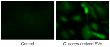
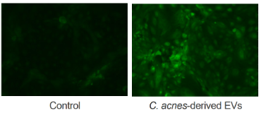
To understand the impact of C. acnes’ extracellular vesicles on keratinocytes or sebocytes, two skin cell types in close contact with the colonizing bacterium.
Our approach at Vibiosphen
- After culture in 1.5L of broth, C. acnes cells were pelleted at 4,500 rpm for 20 min at 4°C and the supernatant was filtered through 0.45 and 0.22 µm vacuum filters. The filtrates were concentrated approximately 20-fold through tangential ultrafiltration using a 100 kDa membrane (Vivaflow system, Sartorius), down to 50 mL. The 50 mL were concentrated through centrifugation on amicon columns using a 50 kDa membrane down to approximately 1 mL.
- The concentration of EVs was estimated using protein concentration determination, using the Bradford assay.
- A fraction of the EVs were labeled with the fluorescent dye Vybrant DiI.
- EVs were added to the culture medium of primary human keratinocytes or sebocytes.
Outcomes of in vitro-Role of microbial-derived extracellular vesicles in pathophysiology
This experimental approach may help to elucidate the role of microbial extracellular vesicles in the pathophysiology of various infections.